


Digital Dentistry

Last updated
Dental technology helps Dr. Michael Kim evaluate oral health, monitor dental development, explain findings to families, and plan pediatric and orthodontic care. Different technologies provide different types of information, so imaging and scanning methods are selected according to each child’s age, symptoms, examination findings, cavity risk, health history, and treatment needs.
Technology supports clinical judgment rather than replacing it. Dental imaging is used only when the expected diagnostic or treatment-planning benefit justifies it.


CBCT
Three-Dimensional CBCT Imaging for Children and Teens in Apex: Cone beam computed tomography, or CBCT, creates detailed three-dimensional images of the teeth, jaws, and surrounding structures. Dr. Kim may recommend CBCT when additional information is needed to evaluate an impacted tooth, tooth development, root position, dental trauma, jaw anatomy, or a complex orthodontic concern.
Because CBCT provides more information and generally involves more radiation than routine two-dimensional dental imaging, it is used selectively. When CBCT is appropriate, the imaging area and exposure settings are selected according to the child’s size and diagnostic needs.
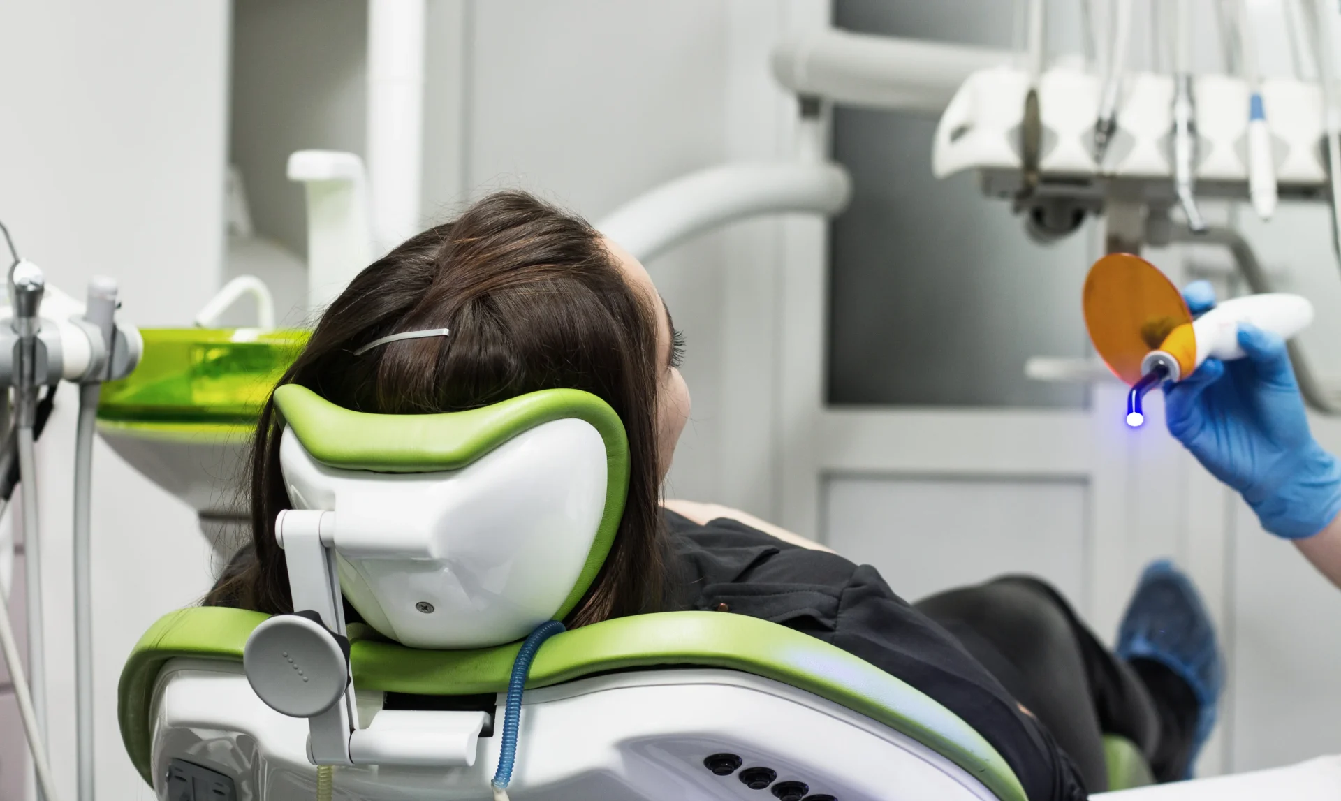

Digital X-rays
Digital Dental X-rays for Children in Apex: Digital X-rays help identify cavities, infections, bone changes, tooth-development concerns, and other conditions that may not be visible during a clinical examination. The images appear quickly on a computer screen, allowing Dr. Kim to enlarge specific areas and explain the findings to parents and caregivers.
The type and frequency of X-rays are based on the child’s age, symptoms, cavity risk, dental development, oral health, and previous images. Children do not receive dental X-rays according to one fixed schedule.

Intraoral Camera
Detailed Intraoral Photography for Children in Apex: An intraoral camera captures enlarged color images of the teeth, gums, and other visible structures inside a child’s mouth. These images can document existing conditions, monitor changes, and help families understand what the dental team sees during an examination.
Intraoral photographs may be useful for showing visible cavities, tooth damage, plaque accumulation, gum concerns, or areas that should be monitored. They complement a clinical examination but do not replace dental X-rays when imaging beneath the surface is needed.

Intraoral Scanner
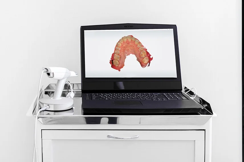
Digital Dental Impressions for Pediatric and Orthodontic Care in Apex: An intraoral scanner captures a series of images to create a three-dimensional digital model of a child’s teeth and bite. Digital scanning may be used when planning braces, clear aligners, retainers, space maintainers, mouthguards, or other customized appliances.
For many children and teens, digital scanning can reduce or eliminate the need for trays filled with conventional impression material. The child must still remain reasonably still while the scanner records the necessary areas.

NOMAD Handheld X-ray
Handheld Digital X-ray Imaging for Children in Apex: A NOMAD handheld X-ray unit allows the dental team to capture digital images while remaining close to the child. Its portable design can support efficient positioning and may be helpful for young patients who need additional instruction or reassurance during imaging.
The dental team follows applicable positioning, shielding, equipment, and radiation-safety protocols whenever handheld imaging is used.
Panoramic X-rays
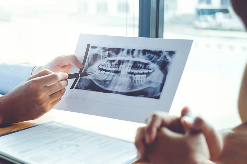
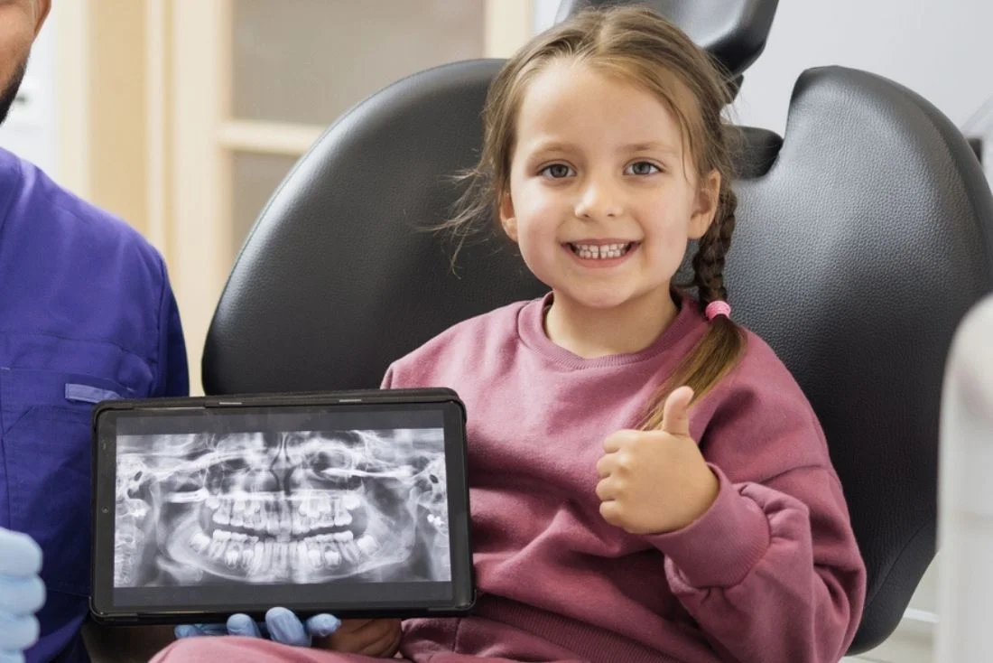
Panoramic Dental Imaging for Children and Teens in Apex: A panoramic X-ray creates a broad, two-dimensional image of the teeth, jaws, jaw joints, and surrounding structures. It may be recommended to evaluate tooth development, eruption patterns, missing or extra teeth, impacted teeth, jaw concerns, or orthodontic treatment needs.
A panoramic image provides a broad overview but does not replace detailed intraoral X-rays or CBCT when another type of imaging is needed for a specific concern.

“Dental technology gives us several ways to evaluate a child’s oral health and dental development. We select each image or scan according to the child’s individual needs so the information collected supports an accurate diagnosis, clear communication with families, and thoughtful treatment planning.”
Dr. Michael Kim
Read Verified Reviews From Apex Parents

“My kids have been going to this practice since they were toddlers, and having one doctor who understands their complete dental history and growth patterns has made such a difference. Dr. Kim treats them like they’re his own children and recommends only what’s truly necessary. The convenience of having both their regular checkups and orthodontics in one place has been a game-changer for our family.”
“What impressed me most was how honest the doctor is about treatment recommendations. He explained exactly why each step was needed and never pushed unnecessary procedures. Having someone I trust completely managing both my kids’ dental and orthodontic care gives me peace of mind. The staff is incredibly kind and patient with nervous children.”
“We switched practices specifically because we wanted someone who could handle both pediatric dentistry and orthodontics under one roof. The continuity of care is amazing, and our kids actually look forward to their appointments. The whole team treats every patient like family, and the clinical expertise is evident in every interaction.”
Research-Reported Benefits of Pediatric Dental Technology
Specificity of Photographic Caries Detection in Children
100%
Sensitivity of Combined Photographs for Primary-Tooth Cavities
99.1%
Accuracy of Combined Photographs for Pediatric Caries Detection
100%
Panoramic Model Sensitivity for Predicting Canine Impaction
90%
Digital X-ray Radiation Reduction Per Exposure
80%
Children Preferring Digital Impressions
75%
0%
10%
20%
30%
40%
50%
60%
70%
80%
90%
100%
0%
50%
100%
| Pediatric Dental Technology Finding | Percentage | What the Research Shows |
|---|---|---|
| Specificity of photographic caries detection | Up to 100% specificity | A JADA study involving children with primary or mixed dentition found that parent-obtained intraoral photographs had specificity ranging from 97.1% to 100% for detecting cavities, depending on the photographic view evaluated. |
| Sensitivity of combined photographs for primary-tooth cavities | Up to 99.1% sensitivity | When photographic views were combined, the study reported sensitivity ranging from 94.8% to 99.1% for detecting cavities in evaluated areas of the primary dentition. |
| Accuracy of combined pediatric intraoral photographs | Up to 100% accuracy | Combined photographs demonstrated accuracy ranging from 94.1% to 100% for the evaluated teeth and photographic views. |
| Panoramic prediction of canine impaction | 90% sensitivity | A prediction model using measurements from panoramic images achieved 90% sensitivity when distinguishing potentially impacted maxillary canines from normally erupting canines. |
| Radiation reduction with digital intraoral imaging | Up to 80% less per exposure | Dental radiology research has reported that digital intraoral receptors may require approximately 50% to 80% less radiation per exposure than conventional film, depending on the system and settings used. |
| Children preferring digital impressions | 75% preferred digital | In a randomized crossover study, 18 of 24 children ages six to 11 preferred digital impressions over conventional alginate impressions. |
Sources: Bharmal RV et al., “A Multicentre Audit to Assess the Effectiveness of the British Orthodontic Society ‘Hold That Smile’ Retainer Videos,” Journal of Orthodontics, 2020; Lee KC et al., “Survival Rates of Mandibular Fixed Retainers: Comparison of a Tube-Type Retainer and Conventional Multistrand Retainers,” Journal of Orofacial Orthopedics, 2024; Johal A, Daly K, and Murray D, “A Comparison of Survival Rates Using 3 Methods of Mandibular Fixed Retainer Fabrication,” American Journal of Orthodontics and Dentofacial Orthopedics, 2025; Laspos C et al., “Survival of Conventionally Bonded Mandibular Retainers With or Without Enamel Sandblasting,” European Journal of Orthodontics, 2023; Forde K et al., “Bonded Versus Vacuum-Formed Retainers: A Randomized Controlled Trial. Part 1,” European Journal of Orthodontics, 2018; American Association of Orthodontists, “Retainers”
These findings come from different technologies, pediatric populations, imaging protocols, and research methods. Intraoral photographs do not replace a clinical examination or necessary dental X-rays. Radiation exposure varies by equipment, settings, image type, patient size, and the area being examined.
Key Findings
Technology Supports Pediatric Care: Digital tools can help evaluate cavities, tooth eruption, jaw development, dental injuries, and orthodontic concerns in children and teens.
Imaging Is Selected Individually: Dr. Kim considers the child’s age, symptoms, dental development, cavity risk, health history, and previous images before recommending X-rays.
Each Image Has a Different Purpose: Intraoral X-rays provide detailed views, panoramic imaging provides a broad overview, and CBCT provides three-dimensional information for selected concerns.
Digital Scanning Supports Orthodontic Planning: Three-dimensional digital models may be used to plan braces, clear aligners, retainers, and other customized appliances.
Images Help Families Understand Care: X-rays and intraoral photographs can be displayed and enlarged so parents and caregivers can better understand examination findings and treatment recommendations.
Radiation Exposure Is Carefully Considered: Imaging is limited to what is clinically necessary, with equipment settings and imaging areas selected for the individual child.
Technology Supporting Your Child’s Care
Schedule an appointment at Green Level Pediatric Dentistry & Orthodontics in Apex. Dr. Michael Kim will determine which examination, imaging, or scanning methods are appropriate based on your child’s oral health, dental development, symptoms, and treatment needs.
Questions Apex Families Ask About Dental Technology
Why might a child need different types of dental X-rays?
Each type of image shows different structures. Intraoral X-rays provide detailed views of individual teeth and supporting bone, panoramic X-rays show a broader view of the jaws, and CBCT provides three-dimensional information for selected diagnostic and treatment-planning needs.
Are digital dental X-rays safe for children?
Digital dental imaging uses controlled radiation exposure and is prescribed only when the expected diagnostic benefit justifies it. The dental team follows current safety recommendations, selects appropriate exposure settings, and limits imaging to the areas needed.
What is the difference between CBCT and a panoramic X-ray?
A panoramic X-ray creates a two-dimensional overview of the teeth and jaws. CBCT produces a three-dimensional dataset that can be viewed from multiple angles. CBCT may be recommended when a two-dimensional image does not provide enough information for a specific concern.
What does an intraoral camera show?
An intraoral camera captures enlarged color images of visible structures inside the mouth. It may show cavities, tooth damage, plaque accumulation, gum concerns, or changes that should be monitored.
Is an intraoral scan the same as an X-ray?
No. An intraoral scanner records the visible surfaces of the teeth and gums to create a digital model. It does not use ionizing radiation or show structures inside the teeth, roots, or supporting bone.
Why might my child need a panoramic X-ray?
A panoramic image may be recommended to evaluate tooth development, eruption patterns, missing or extra teeth, impacted teeth, jaw structures, or orthodontic concerns. Dr. Kim determines whether it is needed based on the child’s age, examination findings, and treatment plan.