


Digital Dental X-Rays for Children and Teens in Apex

Last updated
Digital X-rays help Green Level Pediatric Dentistry & Orthodontics evaluate parts of a child’s teeth and jaws that cannot be seen completely during a clinical examination. These images may be used to identify cavities, monitor dental development, evaluate injuries, and support orthodontic treatment planning.


What Are Digital Dental X-Rays?
Digital dental X-rays use an electronic sensor or phosphor plate instead of conventional film. The resulting images appear electronically and can be enlarged, adjusted, compared with earlier images, and securely stored in the child’s dental record.
Different images serve different purposes. Bitewing X-rays commonly help evaluate cavities between back teeth, while periapical images show an entire tooth and its surrounding bone. Panoramic imaging may be recommended when a broader view of dental development is needed.
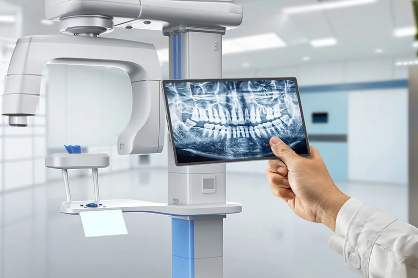
Benefits of Digital X-Rays

Detect Hidden Cavities
Digital X-rays can reveal decay between touching teeth where the area cannot be inspected directly.
Monitor Development
Images can help the dentist evaluate developing permanent teeth, eruption patterns, missing or additional teeth, and available space.
Support Orthodontic Care
Radiographs may provide information about tooth positions, roots, supporting bone, and dental development before or during treatment.
Did You Know…
Children do not receive X-rays solely because they have reached a certain age. The dentist considers the child’s examination, cavity risk, dental development, symptoms, previous images, and treatment needs before prescribing new radiographs.
Ready to Schedule
Your Appointment?

Why Choose Green Level Pediatric Dentistry & Orthodontics?
The Digital X-Ray Process
1
Selecting the Image
Dr. Michael Kim determines which type of X-ray, if any, is needed to answer the clinical question.
2
Capturing the Image
A sensor or imaging plate is positioned for the selected view. The child remains still briefly while the image is captured.
3
Reviewing the Results
The image appears electronically so the dentist can evaluate the area, compare it with previous records, and explain relevant findings to the parent and child.


“Digital X-rays provide information that cannot always be gathered from an examination alone. We prescribe images according to each child’s needs and use them to support early detection, developmental monitoring, and thoughtful treatment planning.”
Dr. Michael Kim
Read Verified Reviews From Apex Parents

“My kids have been going to this practice since they were toddlers, and having one doctor who understands their complete dental history and growth patterns has made such a difference. Dr. Kim treats them like they’re his own children and recommends only what’s truly necessary. The convenience of having both their regular checkups and orthodontics in one place has been a game-changer for our family.”
“What impressed me most was how honest the doctor is about treatment recommendations. He explained exactly why each step was needed and never pushed unnecessary procedures. Having someone I trust completely managing both my kids’ dental and orthodontic care gives me peace of mind. The staff is incredibly kind and patient with nervous children.”
“We switched practices specifically because we wanted someone who could handle both pediatric dentistry and orthodontics under one roof. The continuity of care is amazing, and our kids actually look forward to their appointments. The whole team treats every patient like family, and the clinical expertise is evident in every interaction.”
Research-Reported Findings for Digital Dental Imaging
Specificity for Cavitated Caries in Evaluated Digital Images
96.6%
Average Accuracy for Detecting Proximal Caries in Primary Teeth
95%
Caries-Detection Accuracy With Selected Digital Protocols
94%
Caries-Detection Sensitivity With Selected Digital Protocols
93%
Maximum Reported Radiation Reduction Per Digital Exposure
80%
Minimum Reported Radiation Reduction Per Digital Exposure
50%
0%
10%
20%
30%
40%
50%
60%
70%
80%
90%
100%
0%
50%
100%
| Digital Imaging Finding | Percentage | What the Research Shows |
|---|---|---|
| Specificity for cavitated caries | 96.6% specificity | A review of digital imaging research reported 96.6% specificity for cavitated caries under the evaluated study conditions. |
| Accuracy for proximal caries in primary teeth | Approximately 95% accuracy | A pediatric study reported average accuracy of approximately 95% when identifying proximal caries lesions in primary teeth on selected digital images. |
| Caries-detection accuracy | Up to 94% accuracy | Selected studies have reported accuracy reaching approximately 94% when digital dental images were evaluated under controlled research conditions. |
| Caries-detection sensitivity | Up to 93% sensitivity | Selected digital acquisition and image-processing protocols produced sensitivity reaching approximately 93% for the evaluated caries lesions. |
| Maximum reported radiation reduction | Approximately 80% less | Published dental radiology research has reported that some digital intraoral systems may require approximately 80% less radiation per exposure than conventional film. |
| Minimum reported radiation reduction | Approximately 50% less | The commonly cited reduction range begins at approximately 50%, although the actual difference depends on the receptor, film comparison, equipment, and exposure settings. |
Sources: Berkhout WER, Sanderink GCH, and van der Stelt PF, “Does Digital Radiography Increase the Number of Intraoral Radiographs?” Dentomaxillofacial Radiology, 2003; Uprichard KK et al., “Comparison of Direct Digital and Conventional Radiography for the Detection of Proximal Surface Caries in the Mixed Dentition,” Pediatric Dentistry, 2000; American Dental Association, “X-Rays and Intraoral Imaging”; American Dental Association, “Optimizing Radiation Safety in Dentistry,” Journal of the American Dental Association, 2024; U.S. Food and Drug Administration, “Dental Radiography: Doses and Film Speed”.
Six Key Findings About Digital X-Rays
Different images answer different questions. Bitewing, periapical, occlusal, cephalometric, and panoramic images are not interchangeable.
X-rays can complement a visual examination. Some cavities remain hidden between teeth until they become large enough to produce visible changes.
Previous images may reduce unnecessary exposure. Reviewing existing diagnostic-quality radiographs can help determine whether new images are needed.
Digital enhancement does not replace interpretation. Magnification and contrast adjustments can support evaluation, but the dentist must interpret the complete image in its clinical context.
Two-dimensional imaging has limitations. When overlapping anatomy prevents an adequate evaluation, panoramic imaging or a selectively prescribed CBCT scan may be considered.
Imaging needs change as children grow. Eruption, changing cavity risk, orthodontic treatment, trauma, and new symptoms may affect which images are appropriate.
Schedule a Retainer Appointment in Apex
Families can schedule an appointment with Green Level Pediatric Dentistry & Orthodontics in Apex for a clinical examination and individualized imaging recommendations. Digital X-rays will be prescribed only when they are expected to provide information relevant to the child’s diagnosis or treatment.
Questions Apex Families Ask About Digital X-Rays
What can digital X-rays show in children?
Digital X-rays may reveal cavities between teeth, changes around tooth roots, developing permanent teeth, missing or additional teeth, dental injuries, bone concerns, and eruption problems that cannot be evaluated fully during a visual examination.
Are digital X-rays safe for children?
Dental X-rays use ionizing radiation, so they should be prescribed thoughtfully. The dentist considers the expected diagnostic benefit and uses appropriate equipment, exposure settings, positioning, and image selection for the child.
How often does my child need dental X-rays?
There is no single interval for every child. Imaging frequency depends on cavity risk, symptoms, dental development, previous findings, orthodontic needs, and whether existing images remain diagnostically useful.
Does my child need X-rays before braces or aligners?
Diagnostic images are often part of orthodontic evaluation, but the records needed vary by patient. The dental team selects images according to tooth development, bite concerns, treatment goals, and available previous records.
What if my child has difficulty tolerating the sensor?
The team may use a smaller receptor, adjust its position, offer age-appropriate coaching, or select another suitable imaging method. The goal is to obtain a useful image while making the experience manageable for the child.
Are digital X-rays the same as CBCT?
No. Standard digital X-rays create two-dimensional images of selected areas. CBCT creates a three-dimensional dataset and is reserved for cases in which the additional information is expected to influence diagnosis or treatment.