


Panoramic Dental Imaging for Children and Teens in Apex

Last updated
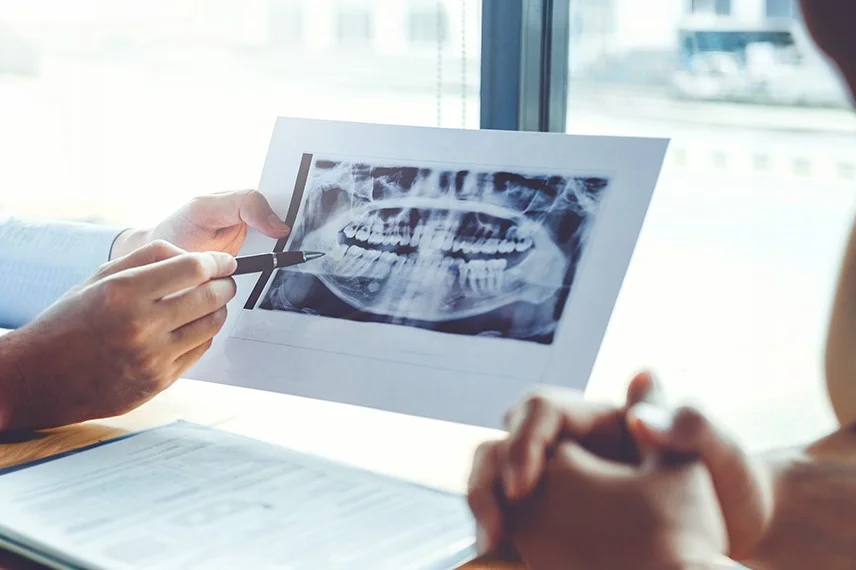
Panoramic X-rays provide a broad, two-dimensional view of a child’s teeth, jaws, and surrounding structures in a single image. Green Level Pediatric Dentistry & Orthodontics may recommend panoramic imaging to evaluate dental development, unerupted teeth, orthodontic concerns, injuries, or selected jaw conditions.


What Is a Panoramic X-Ray?
A panoramic X-ray is taken with an imaging unit that rotates around the child’s head. The resulting image generally includes the upper and lower teeth, developing permanent teeth, jawbones, jaw joints, and portions of nearby anatomical structures.
Unlike bitewing or periapical X-rays, the image receptor remains outside the mouth. The child may bite gently on a positioning guide while remaining still during the brief scan.
Benefits of Panoramic Imaging

Monitors Dental Development
The image can help the dentist evaluate developing permanent teeth, eruption patterns, missing teeth, additional teeth, and retained primary teeth.
Supports Orthodontic Planning
A panoramic X-ray may show general tooth positions, root development, crowding, impacted teeth, and other conditions relevant to orthodontic care.
Provides Broad Coverage
One image captures a larger area than an individual intraoral X-ray, allowing the dentist to review both jaws and the developing dentition together.
Did You Know…
Panoramic images are useful for broad screening and developmental evaluation, but they do not provide the same fine detail as bitewing or periapical X-rays. Additional imaging may be recommended when a particular tooth or structure requires closer evaluation.
Ready to Schedule
Your Appointment?

Why Choose Green Level Pediatric Dentistry & Orthodontics?
The Imaging Process
1
The Imaging Process
The dental team positions the child’s head and may ask them to bite gently on a guide, place the tongue against the roof of the mouth, and remain still.
2
Capturing the Image
The imaging unit rotates around the child’s head. The scan itself is generally completed within a short period.
3
Reviewing the Results
Dr. Michael Kim evaluates the image alongside the child’s examination, dental history, previous images, and orthodontic records.


“A panoramic X-ray gives us a broad view of dental development that cannot be obtained through a visual examination alone. We use it selectively to evaluate eruption, impacted teeth, missing or additional teeth, and other concerns that may affect a child’s treatment plan.”
Dr. Michael Kim
Read Verified Reviews From Apex Parents

“My kids have been going to this practice since they were toddlers, and having one doctor who understands their complete dental history and growth patterns has made such a difference. Dr. Kim treats them like they’re his own children and recommends only what’s truly necessary. The convenience of having both their regular checkups and orthodontics in one place has been a game-changer for our family.”
“What impressed me most was how honest the doctor is about treatment recommendations. He explained exactly why each step was needed and never pushed unnecessary procedures. Having someone I trust completely managing both my kids’ dental and orthodontic care gives me peace of mind. The staff is incredibly kind and patient with nervous children.”
“We switched practices specifically because we wanted someone who could handle both pediatric dentistry and orthodontics under one roof. The continuity of care is amazing, and our kids actually look forward to their appointments. The whole team treats every patient like family, and the clinical expertise is evident in every interaction.”
Research-Reported Diagnostic Findings From Panoramic Images
Discrimination of Impacted From Normally Erupting Canines
97%
Specificity of a Canine-Impaction Prediction Model
94%
Sensitivity of a Canine-Impaction Prediction Model
90%
Specificity for Detecting Congenitally Missing Teeth With Evaluated Software
100%
Negative Predictive Value for Congenitally Missing Teeth
99.4%
Sensitivity for Detecting Congenitally Missing Teeth
Sensitivity for Detecting Congenitally Missing Teeth
0%
10%
20%
30%
40%
50%
60%
70%
80%
90%
100%
0%
50%
100%
| Panoramic Imaging Finding | Percentage | What the Research Shows |
|---|---|---|
| Discrimination of canine impaction | 97% AUC | A prediction model combining three measurements from panoramic radiographs achieved an area under the curve of 0.97 for distinguishing impacted from normally erupting maxillary canines. |
| Specificity for canine impaction | 94% specificity | At the selected cutoff, the model correctly classified 94% of the normally erupting canines evaluated in the study. |
| Sensitivity for canine impaction | 90% sensitivity | The same panoramic model identified 90% of the impacted maxillary canines included in the study. |
| Specificity for congenitally missing teeth | 100% specificity | In a study evaluating diagnostic software on panoramic radiographs, the system correctly classified all evaluated cases without congenitally missing teeth. |
| Negative predictive value for congenitally missing teeth | 99.4% NPV | The evaluated panoramic-image software produced a 99.4% negative predictive value for congenitally missing teeth. |
| Sensitivity for congenitally missing teeth | 84.7% sensitivity | The same study reported 84.7% sensitivity for detecting congenitally missing teeth on the evaluated panoramic images. |
Sources: Alqerban A et al., “Early Prediction of Maxillary Canine Impaction,” Dentomaxillofacial Radiology, 2016; Makrygiannakis MA et al., “Diagnostic Accuracy of an Artificial Intelligence-Based Software in Detecting Dental Anomalies on Panoramic Radiographs,” European Journal of Orthodontics, 2025; Tsolakis AI et al., “Reliability of Different Radiographic Methods for the Localization of Displaced Maxillary Canines,” American Journal of Orthodontics and Dentofacial Orthopedics, 2018; American Dental Association, “X-Rays and Intraoral Imaging”.
The first three percentages apply to a specific mathematical prediction model and should not be interpreted as the accuracy of every panoramic examination. The remaining values concern evaluated diagnostic software rather than unaided clinical interpretation. Panoramic findings must be reviewed by a dental professional and may require intraoral X-rays or CBCT for clarification.
Six Key Findings About Panoramic X-Rays
Panoramic images are two-dimensional. Teeth and anatomical structures may overlap, so the image cannot show every relationship with three-dimensional precision.
Developing teeth can be evaluated before eruption. The image may show permanent teeth forming beneath primary teeth and help assess whether development appears consistent with the child’s age.
The image can help identify missing or additional teeth. Tooth-number abnormalities may influence eruption, spacing, and orthodontic planning.
Panoramic images are not ideal for detecting small cavities. Bitewing or periapical X-rays generally provide greater detail when evaluating individual teeth and areas between teeth.
Positioning affects image quality. Movement, incorrect tongue position, jewelry, and head-positioning errors can blur or distort parts of the image.
CBCT may be needed for selected concerns. If an impacted tooth or nearby root cannot be evaluated adequately in two dimensions, three-dimensional imaging may be considered.
Schedule a Pediatric Dental Evaluation
Families can schedule an appointment with Green Level Pediatric Dentistry & Orthodontics in Apex for an examination and individualized imaging recommendations. Dr. Michael Kim will determine whether a panoramic X-ray is appropriate based on the child’s development, symptoms, previous records, and treatment needs.
Questions Apex Families Ask About Panoramic X-Rays
What does a panoramic X-ray show in children?
It provides a broad view of the upper and lower teeth, developing permanent teeth, jaws, jaw joints, and nearby structures. It may reveal impacted, missing, additional, or unusually positioned teeth.
Why might my child need a panoramic X-ray for orthodontic care?
The image may help evaluate eruption, dental development, crowding, root formation, retained primary teeth, and the general position of unerupted permanent teeth.
Does my child have to hold a sensor inside the mouth?
No. The image receptor remains outside the mouth. The child may need to bite gently on a small positioning guide while the machine rotates around the head.
How long does a panoramic X-ray take?
Image capture is generally brief. Additional time is needed to position the child correctly and explain how to remain still during the scan.
Can a panoramic X-ray find cavities?
It may show some larger areas of decay, but it is not the preferred image for detecting small cavities between teeth. Bitewing X-rays generally provide more detailed cavity information.
Is a panoramic X-ray the same as CBCT?
No. A panoramic X-ray produces one broad two-dimensional image. CBCT produces a three-dimensional dataset and is reserved for situations in which the additional information is expected to affect diagnosis or treatment.